orthokeratinized odontogenic cyst
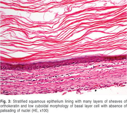
Orthokeratinized odontogenic cyst usually occurs most often between third and fourth and decades and with a male gender predilection711 In this case 30-year-old female was reported with OOC. Histologically OOC is characterized by a 4-8-cell-layer-thick orthokeratinized epithelial lining with prominent granulosum and low cuboidal basal cells.
Jcm Free Full Text Changes In Cellular Regulatory Factors Before And After Decompression Of Odontogenic Keratocysts Html
Background Orthokeratinized odontogenic cyst OOC a newly designated entity of odontogenic cysts is an intraosseous jaw cyst that is entirely or predominantly lined by orthokeratinized squamous e.

. Kshirsagar K Shah S Kheur S. They were first identified by Wright in 1981 2 and were originally thought to be part of the spectrum of Odontogenic Keratocyst OKC 3. Background Orthokeratinized Odontogenic Cyst OOC is a rare developmental odontogenic cyst which was considered in the past to be a variant of Odontogenic keratocyst OKC later renamed as.
J Dent Res Prac 2019. The orthokeratinized odontogenic cyst OOC is a rare developmental odontogenic cyst that has been considered as a variant of the keratocystic odontogenic tumour KCOT until Wright 1981 defined it as a different entity. Multiple Orthokeratinized Odontogenic Cysts.
A report of two cases and review of the literature. While KCOT epithelial lining is thick parakeratinized with the basal cells exhibiting typical palisading of the nuclei. Orthokeratinized odontogenic cyse developmental odontogenic cysts characterised by an orthokeratinized strati- fied squamous epithelial lining.
On comparing the OOC with the other developmental cyst particularly dentigerous cyst and OKC it shows a typical distinguishing clinicopathological feature. OOC was actually first described as a dermoid cyst as far back as 1927 by Schultz 8. Orthokeratinized odontogenic keratocyst crossing mandibular midline.
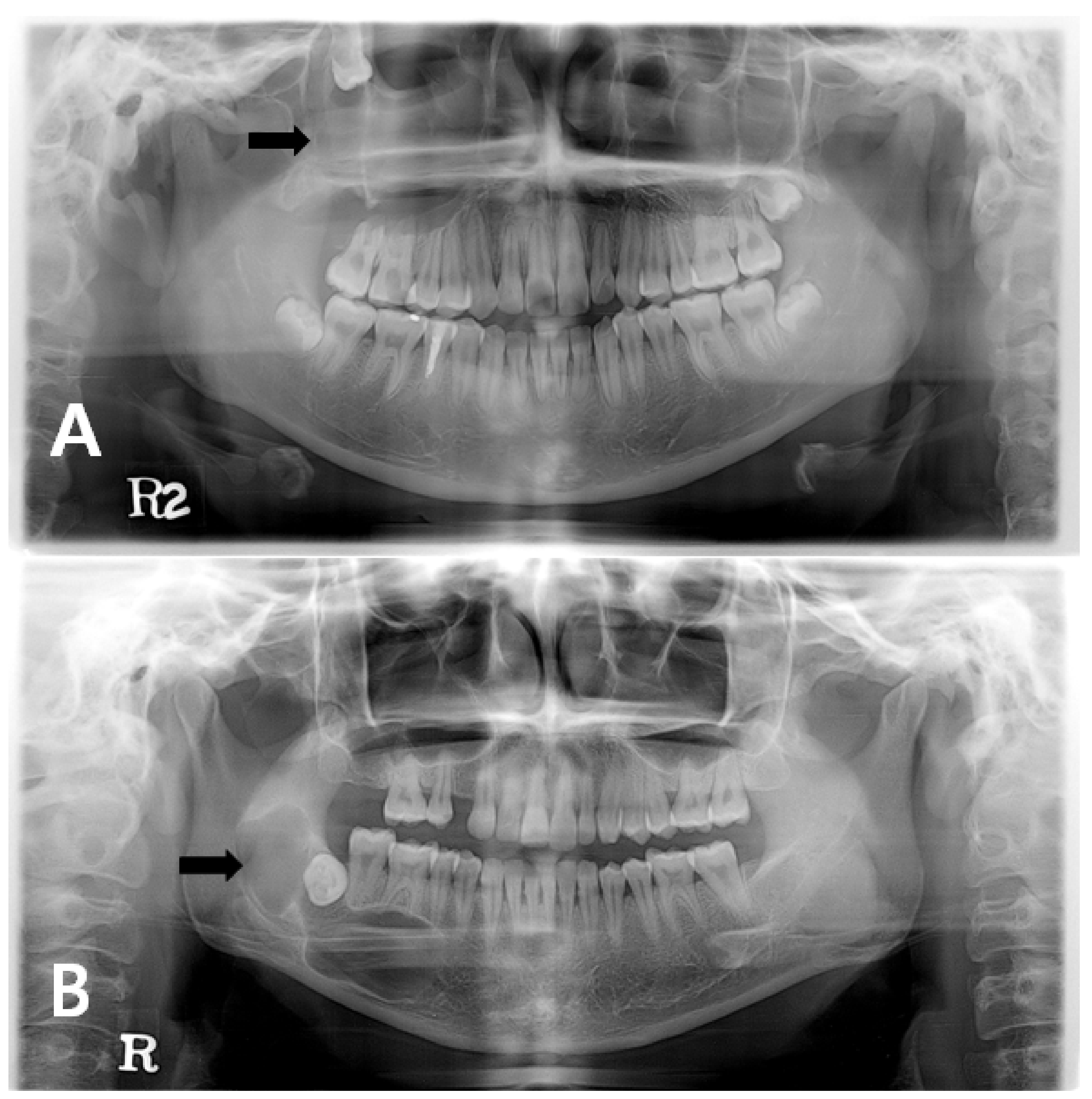
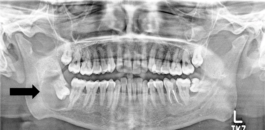
The orthokeratinized odontogenic cyst OOC is a rare developmental odontogenic cyst that has been considered as a variant of the keratocystic odontogenic tumour KCOT until Wright 1981 defined it as a different entity. In contrast to other studies the lesion was located in the body of mandible starting from lower left canine region involving lower left premolars and lower left first molar. OOC had been previously coded as odontogenic keratocyst OKC and was termed as orthokeratinized variant of OKC 2 3 4.
Orthokeratinized Odontogenic Cyst OOC is a developmental odontogenic cyst characterised by a lining of orthokeratinized stratified squamous epithelium 1. In this case 30-year-old female was reported with OOC. OOCs were first described in 1927 by Schultz 2 as a variant of odontogenic keratocysts now known as keratocystic odontogenic tumours KCOTs 3.
The keratocystic odontogenic tumor which was previously referred to as odontogenic keratocyst must be differentiated from other odontogenic cysts because of its aggressive behavior. Orthokeratinized odontogenic cyst OOC is a rare intraosseous cyst characterized by an orthokeratinized epithelial lining and minimal clinical aggressiveness 1. Odontogenic keratocyst was designated as keratocystic odontogenic tumor KCOT in the new World Health Organization classification and OOC should be distinguished from KCOT for.
Orthokeratinized odontogenic cyst OOC was first described by Schultz in 1927 and in 1945 Philipsen considered it to be a variant of Odontogenic keratocyst OKC. Orthokeratinized odontogenic cyst OOC is a relatively uncommon developmental cyst comprising about 10 of cases that had been previously coded as odontogenic keratocysts. In contrast to other studies the lesion was located in the body of mandible starting from lower left canine region involving lower left premolars and lower left first molar.
It is a fairly unusual devel-opmental cyst comprising of merely 04 of odontogenic cyst. OOC exhibits distinctive clinical pathologic and behavioral features that varied substantially from KCOT and hence now it is considered as a separate entity. Orthokeratinized odontogenic cyst OOC is a rare developmental odontogenic cyst characterized by orthokeratinized stratified squamous epithelial lining 1.
Keratocystic odontogenic tumors can be seen at any age but are most common between 10 and 40 years of age in males and within the posterior mandible. Discussion Orthokeratinized Odontogenic Cyst was included as a separate and specific entity for the first time in the 4th Edition of the World Health Organization WHO Classification of Head and Neck Tumors which was published in 2017 7. Orthokeratinized odontogenic cyst usually occurs most often between third and fourth and decades and with a male gender predilection.
Orthokeratinized odontogenic cyst OOC is a sporadic developmental odontogenic cyst. They were originally believed to be part of the spectrum of Odontogenic Keratocyst but are now considered to be a distinct entity. Surgery is the usual treatment and recurrence or association with Gorlin-Goltz syndrome has rarely been described.
Surgery is the usual treatment and recurrence or association with Gorlin-Goltz syndrome has rarely been described.

Odontogenic Cysts Pocket Dentistry

Pin On Oral Pathology

Pin On Oral Pathology

A Case Report And Literature Review Of Multiple Orthokeratinizing Odontogenic Cysts The Great Mimicker Joseph 2021 Oral Surgery Wiley Online Library
Pin By Neha On Oral Pathology Oral Pathology Dentistry Pathology

Pdf Orthokeratinized Odontogenic Cyst A Case Report A Milder Variant Of Okc Or An Independent Entity Semantic Scholar

Odontogenic Keratocyst Oral Pathology Pathology Oral

Pin Page
Pathology Outlines Orthokeratinized Odontogenic Cyst

The Photomicrograph Shows Cystic Lumen Lined By A Continuous Layer Of Download Scientific Diagram

Orthokeratinized Odontogenic Cyst

Pdf Orthokeratinized Odontogenic Cyst A Report Of Two Cases In The Mandible
Orthokeratinized Odontogenic Cyst Critical Appraisal Of A Distinct Entity

Pdf A Cbct Report Review Emphasizing The Characteristics Of Orthokeratinized Odontogenic Cyst Semantic Scholar

Orthokeratinized Odontogenic Cyst Stratified Squamous Epithelial Download Scientific Diagram
2

Pin On Oral Pathology
Pdf Orthokeratinized Odontogenic Cyst

Figure 2 Orthokeratinized Odontogenic Cyst A Report Of Three Clinical Cases


Comments
Post a Comment